Our Facilities
We developed our own in-house built custom equipment that uses state of the art components.
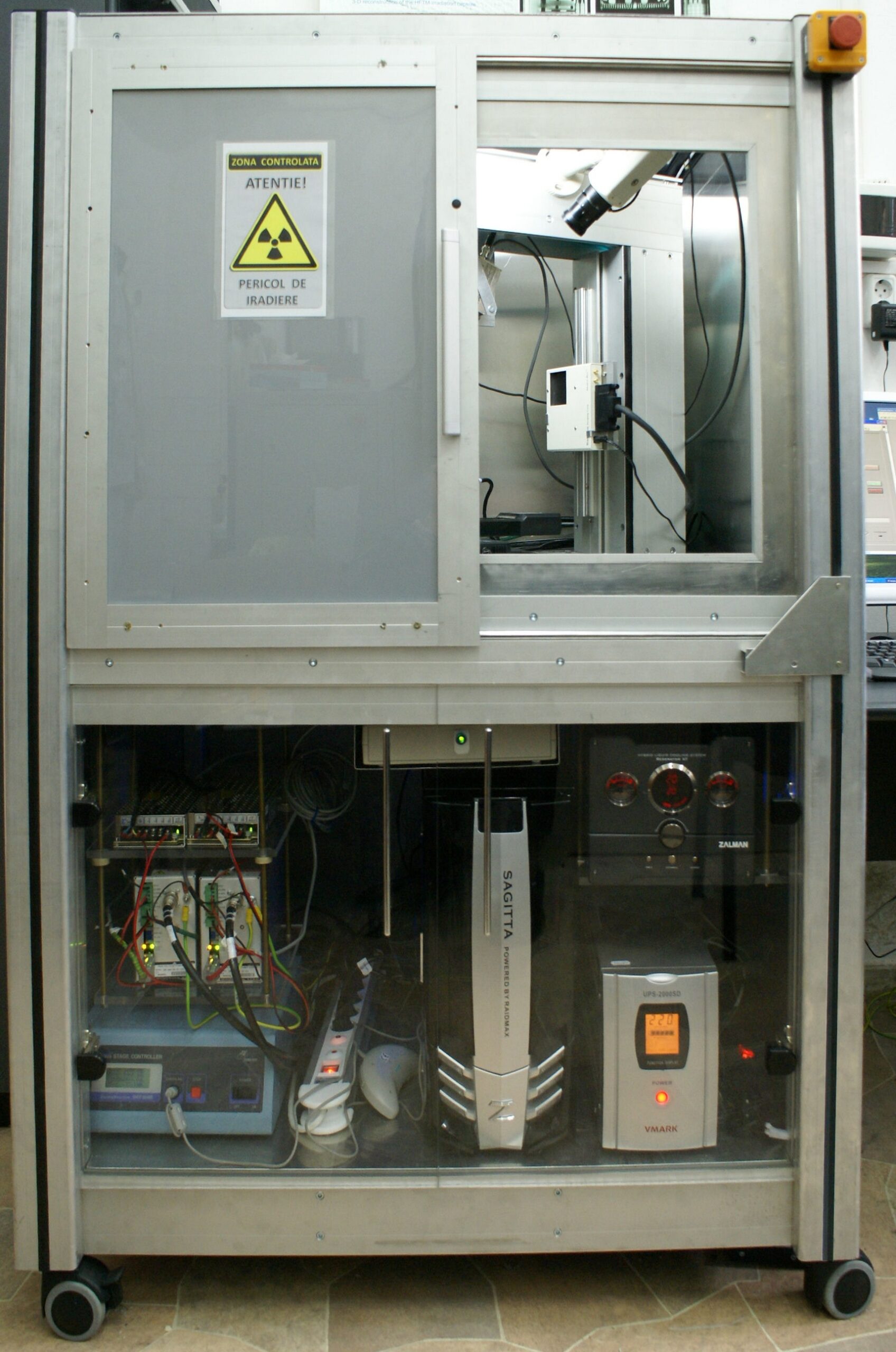
A custom designed versatile sub-micron tomograph capable of achieving resolutions down to 0.9 µm. That value is the minimum size of individual features that can be visualized / analyzed within the internal structure of objects. A wide range of samples types and sizes can be analyzed using different X ray detector types for various custom experimental setups. In-house developed software.
· X ray generator: nano-focus transmission operating at max. 225 KV
· X-ray detector: flat panel 400 x 400 mm, 4000 x 4000 px, pixel size 100 µm, 16 bit
· Seven micrometric motorized axis.
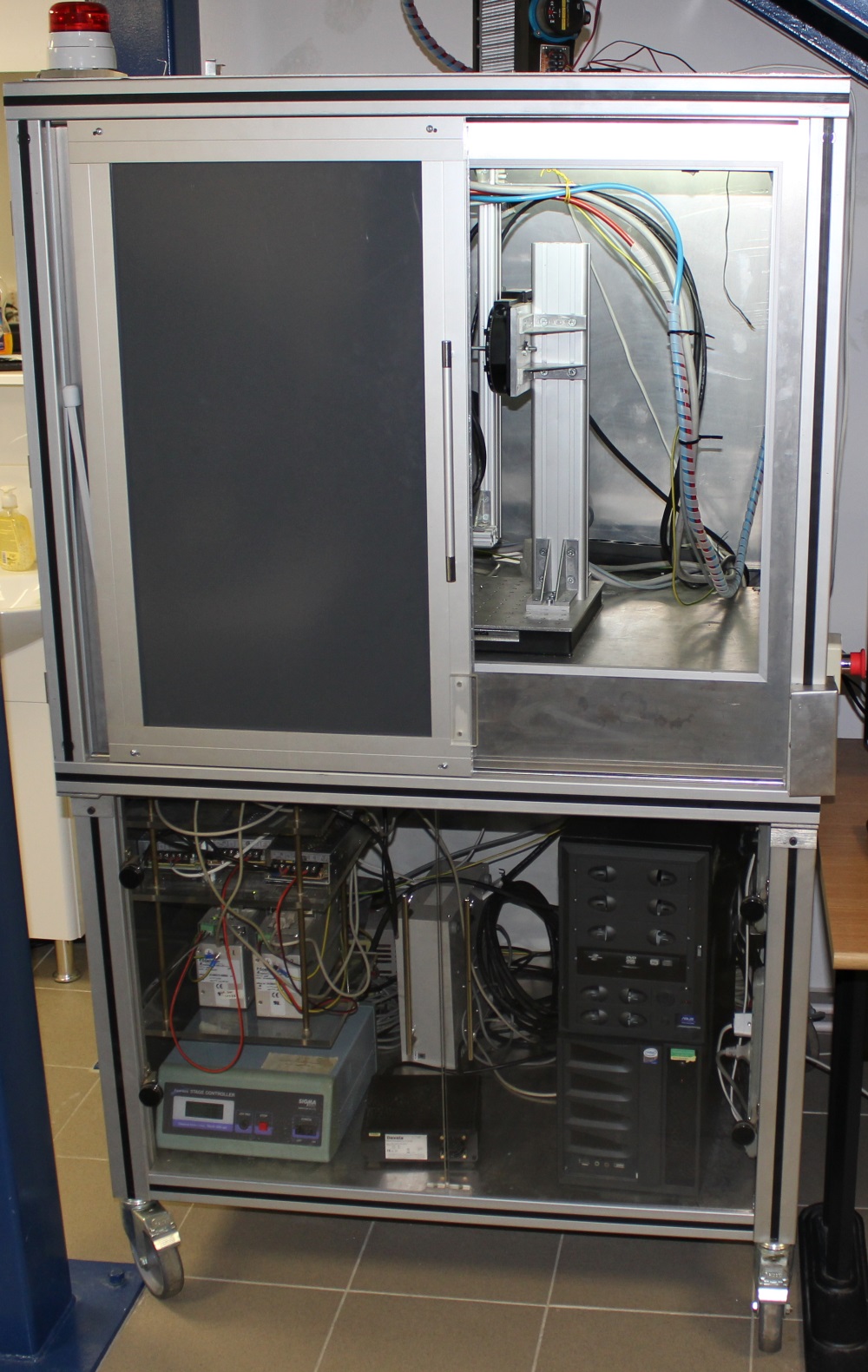
A high-power micro-tomograph designed and developed in-house that can provide high penetrability and resolutions down to 3 µm. The high energy X ray generator allows for a wide range of samples types, densities and sizes to be analyzed. Different X ray detector types are available for various custom experimental setups. We have developed different scanning protocols for samples that can be as small as tens of micrometers or longer than one meter.
- X ray generator: micro-focus with 225 KV & 320 KV interchangeable modules;
- X ray detector: 400 x 400 mm flat panel, 2000 x 2000 px, pixel size 200 µm, 16 bit;
- Four ultra-high precision motorized stages;
- Four high load, high precision motorized stages.
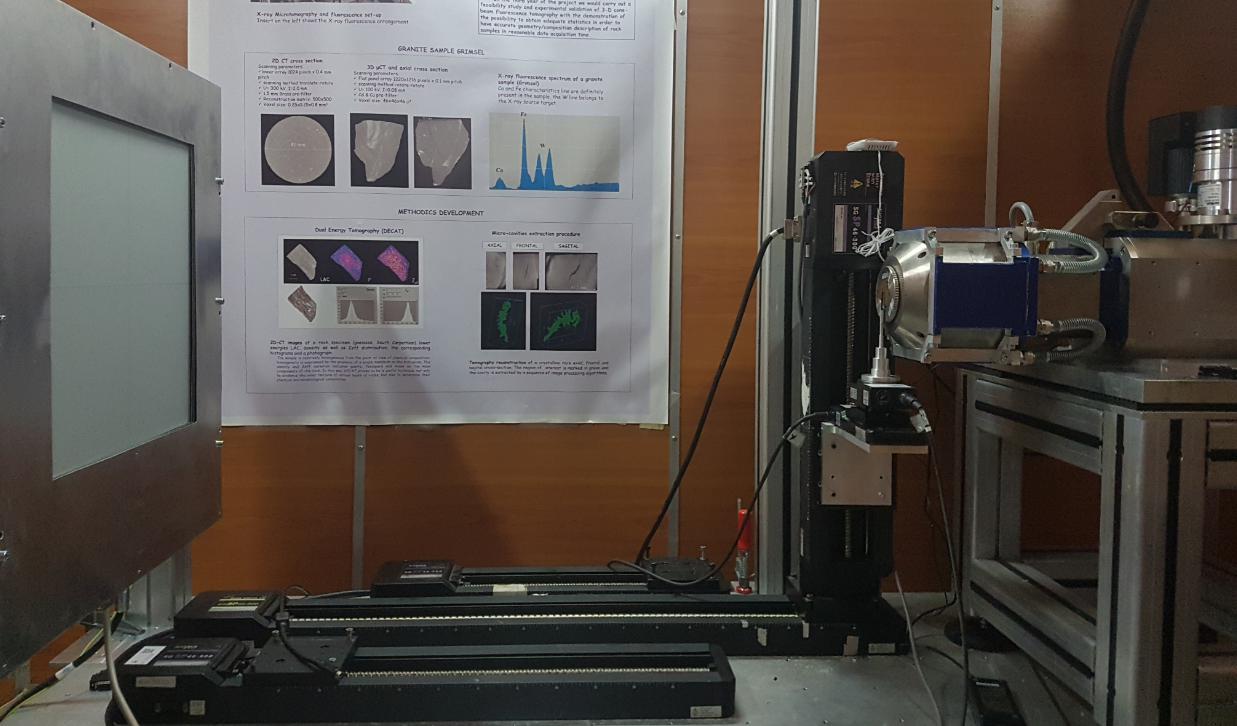
An in-house developed instrument that combines cone-beam tomography (3D-CT) and X-ray fluorescence (XRF) in one compact, modular, versatile, mobile system for noninvasive 3-D morphology and composition mapping.
The 3D micro-tomography is used for microstructural characterization of samples. Internal sample features of few tens of micrometers can be clearly distinguished. The fluorescence component can provide local qualitative and quantitative information about the sample composition elements.
Microtomography setup:
– X-Ray microfocus tube – W anode, 50 KV, 50W
– X-ray flat panel detector – 1024 x 1024 px, pixel size 48 µm, 12 bit
Microfluorescence setup:
– X-Ray microfocus tube – Mo anode, 50 KV, 50W
– Policapilary lens for focusing the output of X-rays
– X-ray miniaturized spectrometer.
Micrometric manipulator with 4 ultra-high precision axis
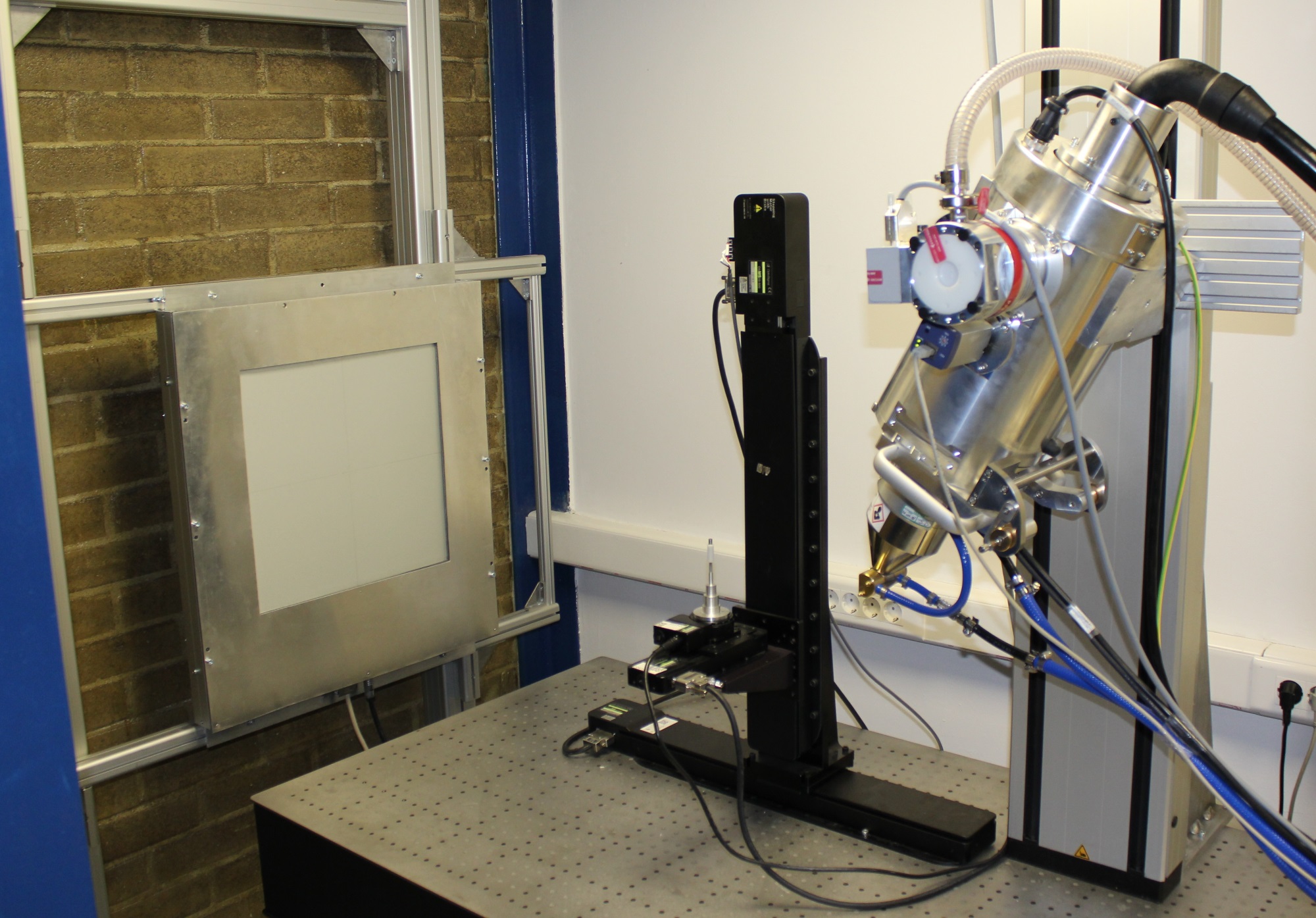
A mobile in-house built modular X-ray system used primarily as a gantry cone-beam tomograph for biology (small animals, plant roots, seeds etc.) and process tomography applications (manufacturing / near conveyor belt, environmental research, corrosion analysis, filters development, polymerization, seed germination, fruit dehydration, etc.).
It features two modules, a frame for performing gantry tomography and a frame that can accommodate the equipment for performing regular X ray microbeam-fluorescence (µXRF). This multifunctional X-ray system can be used for both noninvasive 3-D morphology and composition mapping.
Module 1 – Gantry tomography setup:
– X-Ray microfocus tube – W anode, 50 KV, 50W
– X-ray flat panel detector – 1944 x 1536 px, pixel size 75 µm, 12 bit
Module 2 – Microfluorescence setup:
– X-Ray microfocus tube – Ag anode, 50 KV, 50W
– Policapilary lens for focusing the output of X-rays
– X-ray miniaturized spectrometer
Micrometric manipulator with 4 ultra-high precision axis
Let's do great things together!
We can help with a custom design fitted for your particular application.
Address
Str. Atomistilor 409, 077125, Bucharest-Magurele, Romania
Call Us
+40 21 4574051