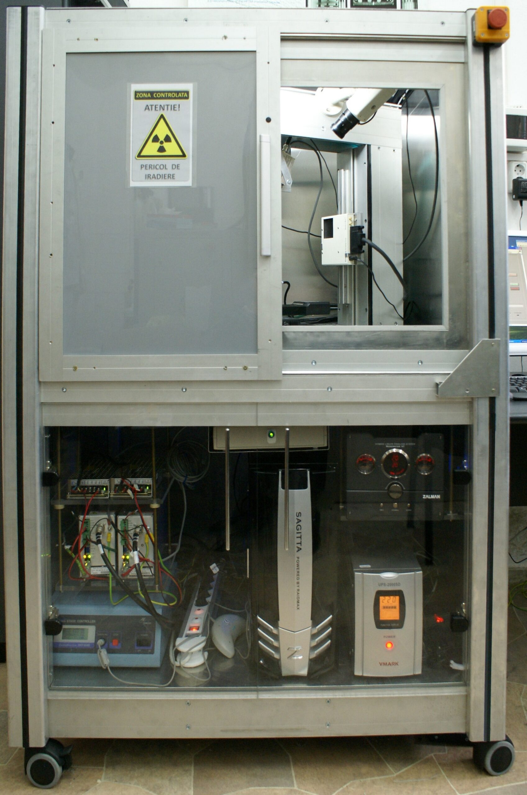
Tomo-Analytic - Compact Mobile X-ray Tomography & Fluorescence System
This is an in-house developed instrument that combines cone-beam tomography (3D-CT) and X-ray fluorescence (XRF) in one compact, modular, versatile, mobile system for noninvasive 3-D morphology and composition mapping.
The 3D micro-tomography is used for microstructural characterization of samples. Internal sample features of few tens of micrometers can be clearly distinguished. The fluorescence component can provide local qualitative and quantitative information about the sample composition elements. Using the X-Y linear stages, the µXRF system provides high resolution (~20 µm) composition mapping and accurate thickness measurements of multilayer samples.
X-ray microtomography is a non-destructive inspection technique that enables the visualization of the internal structure of the samples down to details of few microns. The method requires the acquisition of a large number of images of the sample from different angles, in order to accurately reconstruct its 3D model. Samples can be characterized morphologically and compositionally by visualizing the variations in density in volume, pores, microdefects, with the possibility of performing high-precision 3D geometric measurements.
X-ray microfluorescence is a non-destructive process of spectral inspection and analysis by which the constituent chemical elements of the samples can be determined both qualitatively and quantitatively. This type of analysis is also applicable in determining the thickness of thin layers of objects whose qualities are improved by metal deposits on their surface, both for the research-development field and for the industrial field. The method allows high spatial resolution mapping of the composition and thickness of the coatings of these samples.
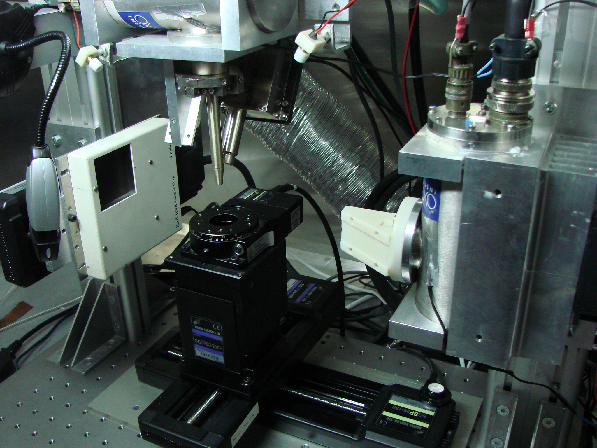
Microtomography setup:
- X-Ray microfocus tube – W anode, 50 KV, 50W,
- X-ray flat panel detector – 1024 x 1024 px, pixel size 48 µm, 12 bit
Microfluorescence setup:
- X-Ray microfocus tube – Mo anode, 50 KV, 50W
- Policapilary lens for focusing the output of X-rays
- X-ray miniaturized spectrometer.
Micrometric manipulator with 4 axis: X – 200 mm,
Y – 200 mm, Z – 20 mm, Rot – 360o
Cabinet exterior size: footprint 1000 mm x 850 mm,
height 1500 mm
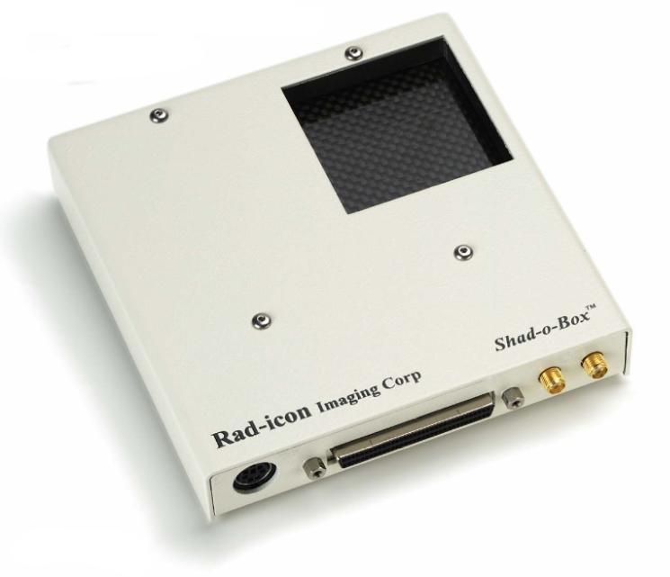
Available X-ray detectors:
Compact small pixel flat panel
Technical Specifications:
– Active area: 410 x 410 mm; Resolution: 1024 x 1024 pixels;
– Pixel size: 48 μm; 12-bit;
– Suitable for small samples with intricate internal structure
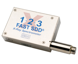
Fast SDD Spectrometer
Technical Specifications:
– Silicon Drift Detector (SDD) with CMOS preamplifier
– Resolution: 122 eV FWHM at 5.9 keV
– Compact size; Suitable for low-energy detectable materials
Address
Str. Atomistilor 409, 077125, Bucharest-Magurele, Romania
Call Us
+40 21 4574051